
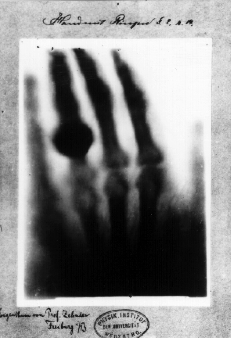
In 1895, the average person traveled by horse-drawn carriage, relied on gas lamps for light, and communicated via telephone. This milestone year also marks the discovery of the X-ray.
Now fast forward 130 years. We’ve evolved to driving cars, lighting our way with electricity, and video chatting over high-speed WiFi. Yet the X-ray is still one of the most widely used diagnostic tools in modern medicine for these reasons:
- It’s non-invasive.
- It’s effective for visualizing bones and other internal structures.
Despite its current applicability, methods for radiographic imaging are stuck in the past. It’s time for a change.
Limits and Ramifications of Traditional X-ray Imaging
Even though radiographic imaging is still instrumental in modern medicine, the equipment used is inefficient.
Operational Inefficiencies
Legacy X-ray systems are expensive, large, immobile, and hard to use. They take significant time to set up and often require several reshoots and patient repositioning to capture the necessary images.
Because you can’t separate the pieces of devices such as C arms and mini C arms, the entire machine must be moved for every adjustment, which can be time-consuming. These inefficiencies can lead to workflow disruptions, increased patient wait times, and slow down healthcare providers’ ability to provide prompt diagnosis.
Facility Constraints
Traditional X-ray machines typically require dedicated, lead-lined rooms to operate safely. This restriction limits access to imaging, meaning orthopedic surgeons can’t capture X-rays onsite and instead send patients to specialized radiology suites when imaging is needed.
Orthopedic surgeons already struggle to keep up with patient volumes, and the situation is exacerbated when they rely on radiology suites for imaging rather than handling it at the point of need.
Meanwhile, patients are shuffled between doctors’ offices, radiology centers, and imaging centers. Each step in the process means another appointment, another commute, and another patient check-in, slowing down crucial diagnoses and treatments.
Staffing Shortages
With legacy radiographic imaging technologies, trained specialists are needed in two key areas:
- To operate the equipment and capture X-rays (radiology technicians, or rad techs)
- To interpret the images and make diagnoses (radiologists)
We’re experiencing a global shortage of both rad techs and radiologists, while our aging population is driving an increased demand for medical imaging.
The American Society of Radiologic Technologists (ASRT) recently conducted a survey revealing an 18.1% vacancy rate of rad techs, up from 6.2% in 2021 (2).
Meanwhile, radiologists are also in short supply; they’re cited as one of the most burnt-out specialists in US healthcare, and fewer graduates are choosing a radiology path coming out of school. The profession is essentially aging out, and the trend doesn’t appear to be changing course any time soon. The Neiman Health Policy Institute reported in February 2025 that the imbalance between the supply of radiologists and the demand for medical imaging will persist until 2055 (3).
These staff shortages amplify the accessibility challenges of radiographic imaging, preventing real-time imaging and forcing healthcare providers to make treatment decisions without immediate visual confirmation.
Combined, operational inefficiencies, facility constraints, and staffing shortages are leaving the medical imaging field in crisis. Many healthcare providers restrict their use of X-rays to highly qualified cases. This means that some injuries and conditions that could benefit from imaging go undiagnosed or are diagnosed later than necessary. For orthopedic surgeons, delayed imaging can lead to prolonged patient discomfort, delayed recovery, and suboptimal treatment decisions.
A New Era: Dynamic Digital Radiography and Portable Imaging Solutions
The radiographic imaging industry is on the cusp of a significant transformation, driven by advances in imaging techniques and mobile technology. One of the most promising innovations is Dynamic Digital Radiography (DDR), a new approach that enhances traditional X-ray capabilities by combining motion analysis with reduced radiation exposure.
DDR captures continuous sequences of images over time, enabling a deeper understanding of joint movement, lung function, and musculoskeletal conditions. While fluoroscopy is still preferable for real-time use cases, such as image-guided injections, DDR provides superior contrast and image clarity, for example, when evaluating joint instability or other applications where the image doesn’t need to be delivered in real time.
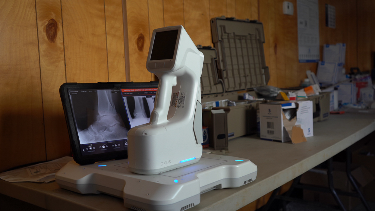
At the same time, breakthroughs in portable X-ray technology, such as OXOS Medical’s MC2 device, are addressing the mobility and accessibility challenges of legacy equipment. Compact, easy to operate, and designed for use in non-traditional settings, these solutions bring high-quality imaging directly to the point of care, whether in a clinic, operating room, or even a sports field.
Faster Diagnoses, Better Patient Outcomes
By integrating DDR and portable imaging solutions into routine care, orthopedic surgeons and other specialists can:
- Diagnose injuries in real time, without waiting for the radiology department’s availability.
- Perform image-guided procedures with greater precision and reduced radiation exposure.
- Expand access to imaging in underserved areas or high-demand settings like emergency rooms and sports medicine clinics.
The Time for Change is Now
The radiographic imaging industry can no longer afford to operate under outdated constraints. The combination of DDR and portable X-ray technology offers an opportunity to revolutionize how imaging is used in healthcare. By embracing these advancements, medical professionals can deliver faster, safer, and more efficient care, improving patient outcomes and redefining standards in diagnostic imaging.
The transformation of radiographic imaging is no longer a distant vision; it is happening now. With innovative solutions like the MC2, healthcare providers can overcome long-standing barriers, making high-quality X-ray imaging a standard part of everyday patient care rather than a limited, inconvenient resource.
The future of radiographic imaging is mobile, efficient, and seamlessly integrated into clinical workflows. By embracing this shift, the industry can enhance accessibility, improve patient outcomes, and usher in a new era of diagnostic excellence.
Resources
(1) https://www.ncbi.nlm.nih.gov/books/NBK546155/
(2) https://www.rsna.org/news/2024/october/radiologic-technologist-shortage