
Capturing X-rays of the hand can be, well, a handful – in more ways than one.
In terms of volume, hand X-rays are in high demand. Meanwhile, it can be challenging to capture quality images of the hand. Legacy systems can be cumbersome and complicated to operate, while the hand’s anatomy adds complexities and intricacies to the process. Bulky machines, restrictive environments, and awkward patient positioning slow clinicians down when speed and precision matter most.
But there is good news: the MC2 portable X-ray device from OXOS is changing this. Hand surgeons can incorporate hand X-rays seamlessly into their care regimens with MC2, designed for faster diagnosis and better patient outcomes.
The High Demand for Hand X-rays
About 25% of the human body’s bones are found in the hand, and we use our hands for just about every task in everyday life. It makes sense that X-rays of the hand are among the most frequently ordered imaging studies in orthopedic care.
Hand X-rays are routinely ordered for:
- Fractures and dislocations
- Arthritis and degenerative changes
- Ligament or tendon injuries
- Post-surgical evaluation
- Occupational or sports-related injuries
Whether to evaluate a wrist fracture or track post-op healing, the ability to capture high quality imaging of the hand is a critical component of diagnosis and treatment planning in emergency medicine, orthopedics, urgent care, and primary care.
4 Reasons the Status Quo Can’t Keep Up
Even though hand X-rays are a mainstay of most modern orthopedic practices, the technologies that most providers rely on for radiographic imaging are stuck in the past and mired in inefficiency. There are four major drawbacks of existing approaches to hand X-rays.
Traditional X-ray machines are large, fixed devices. It’s not easy to move them between different care settings or get them into the appropriate position to capture unique angles and small anatomy, which is a big part of capturing X-rays of the hand. The hand’s anatomy is small, intricate, and – especially in cases involving trauma – it can be difficult to get into the right position for a quality image at the right angle(s).
Legacy machines are complex to operate, requiring radiologic technologists to capture the images, which are then sent to radiologists, orthopedic surgeons, or other specialists for interpretation. With the industry shortage of both rad techs and radiologists, they’ve become a bottleneck, limiting throughput of the radiology department and impacting the quality of care patients receive. The ability to see what’s really happening in the patient’s hand depends on technician availability. There’s pressure to focus rad techs’ and radiologists’ time on higher value work than capturing and interpreting hand X-rays.
Traditional X-ray machines are confined to lead-lined rooms to limit their radiation emission. This further inhibits the accessibility of X-rays to facilities with lead-lined rooms, adding another dependency to the patient’s ability to get an X-ray of the hand – another appointment requiring scheduling and transportation.
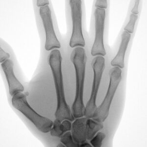
Image quality is of paramount importance with X-rays of the hand. The hand contains 27 small bones (including phalanges, metacarpals, and carpals), along with joints, tendons, ligaments, and growth plates. These structures are densely packed, and pathology (e.g. a hairline fracture or early arthritis) can be extremely subtle. (Compare this, for example, to a chest or pelvis X-ray, where anatomy is larger and more easily distinguished.) The hand uniquely demands finer spatial resolution to identify small-scale abnormalities and subtle findings such as hairline fractures, early arthritic changes, or growth plate shifts.
Introducing MC2: A New Approach to X-rays of the Hand
MC2 is the world’s first smart, portable, and cordless digital X-ray system for static and dynamic imaging of extremities. FDA-cleared for use without a lead-lined room, MC2 helps healthcare practitioners improve patient outcomes at the point of need, wherever that may be. MC2 is easy, versatile, and accessible – making it ideal for X-rays of the hand.
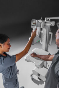
MC2 is easy to operate. The portable X-ray device is designed for efficiency, streamlining the process for patients and care providers. Its simple interface appeals to a wider user base of medical practitioners and has a short learning curve for clinical staff who are new to X-ray technologies. MC2 is easy enough to be operated with one hand or hands-free, freeing clinicians to help position the patients’ intricate hand anatomy.
For instance, MC2 includes an integrated viewfinder to help properly align anatomy, along with built-in safeguards such as its patented “no-fire” positioning system which determines the distance and angle from X-ray source to detector. This prevents users from emitting radiation unless the device is properly aligned. And MC2 includes an automatic collimator to limit X-ray beam field size and collimation pucks for additional manual adjustment.
MC2 is versatile to suit the needs of hand X-rays, with excellent image clarity and accuracy, modern automations, and flexible imaging configurations.
MC2’s compact size, ergonomic design, and accessories accommodate different anatomical positions and help operators capture off-angle shots when needed.
MC2 is accessible. It boasts a portable form factor and low radiation profile – 40-80 kV tube voltage and radiation intensity of 1-2 mA, yielding a small scatter area.
This makes MC2 easy to integrate within diverse care settings and workplaces, including within orthopedic practices, urgent care facilities, and sports medicine environments, where hand injuries are frequent and speed matters. No more delays, piecemeal appointments, or hand-offs.
Use Cases Across 4 Imaging Modes
The MC2 portable X-ray device offers radiographic diagnostics via four distinct imaging modes – radiography, dynamic digital radiography (DDR), fluoroscopy, and photography – all of which deliver the high image resolution and precision that are important for small bone structures in the hand. Here are example use cases for the hand across each imaging mode:
- Standard Radiography is used to diagnose fractures, arthritis, or growth plate changes with crisp, static images.
- Dynamic Digital Radiography (DDR) captures joint motion which is ideal for evaluating wrist instability, ligament injuries, or pre/post-op joint function.
- Fluoroscopy offers real-time visualization, which is critical for image-guided injections or foreign body removal.
- Photography Mode helps with visual documentation of soft tissue injuries for surgical planning or progress tracking.
See MC2 in Action
With MC2, orthopedic surgeons are transforming X-rays of the hand with quality and streamlined processing.
Want to see how the MC2 fits into your practice? Request a demo and experience the future of orthopedic imaging firsthand (pun intended).