

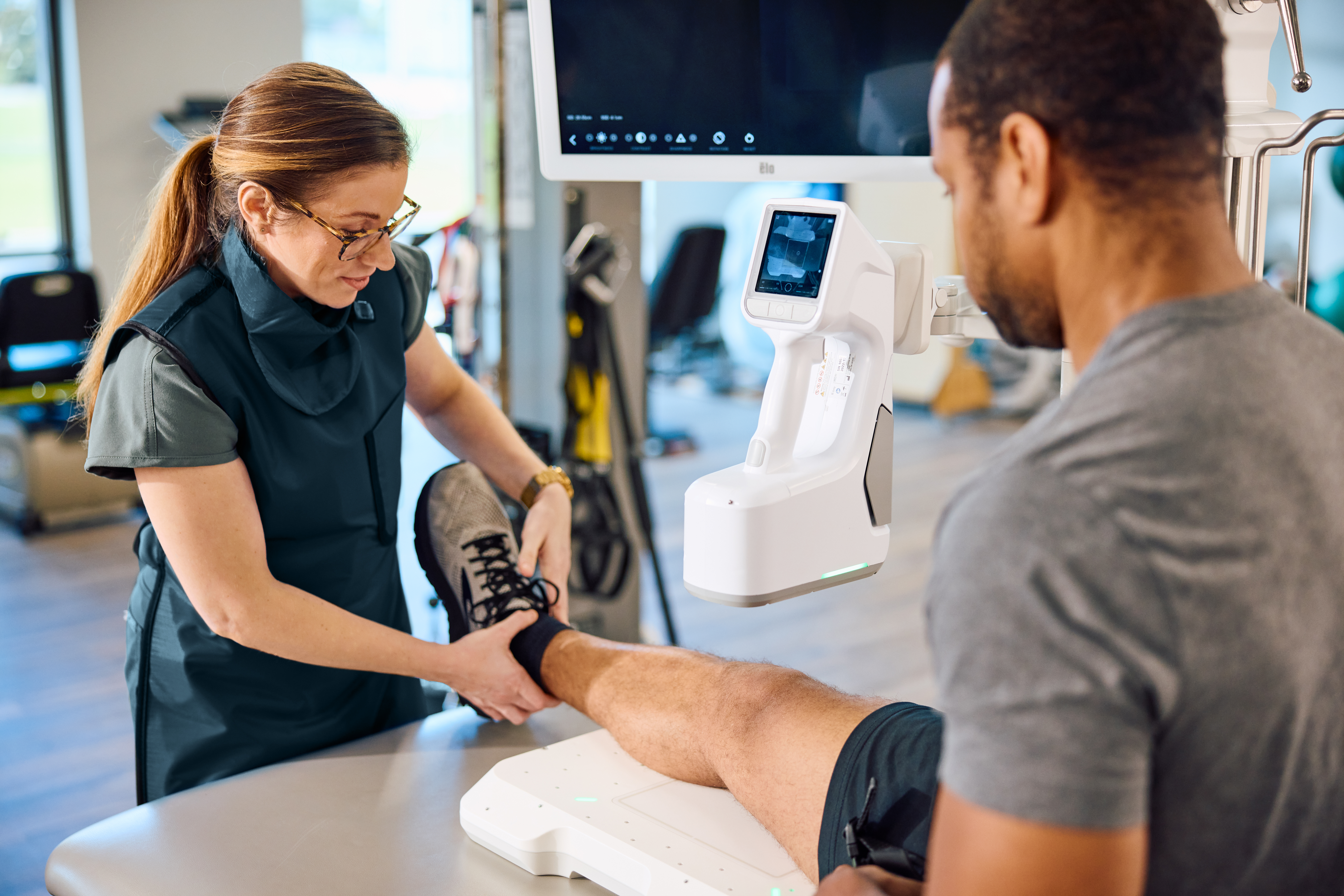
The knee is the largest joint in the human body, and it quite literally carries a lot of weight. The simple act of walking downstairs can load the knee with three to five times a person’s body weight; it’s no surprise that knees are among the most frequently imaged joints in orthopedic care.
Clinically, the reasons for imaging the knee are wide-ranging. X-rays are routinely ordered for patients presenting with trauma, pain, swelling, or an inability to bear weight, as well as for evaluating osteoarthritis and other degenerative changes. As a result, knee imaging is a daily, high-volume task in orthopedic clinics, emergency departments, and specialty practices.
Yet, despite how often knee X-rays are called for, the process for capturing them leaves much room for improvement. Traditional X-ray systems can make knee imaging feel awkward, inefficient, and uncomfortable. For a joint that’s imaged often and under varied conditions, a more flexible approach is needed.
But before we get into that, let’s quickly recap the many situations that warrant knee X-rays.
Many Care Scenarios Call for Knee X-rays
Knee X-rays are an effective way to diagnose and treat the high-value joint across a broad spectrum of clinical scenarios, from urgent injury assessment to longer-term joint evaluation. In acute settings, imaging is used to investigate suspected fractures or other traumatic injuries, particularly when patients are unable to bear weight or tolerate significant movement.
Beyond trauma, knee X-rays are routinely used to evaluate chronic and degenerative conditions. For patients with suspected or known osteoarthritis (OA), imaging helps assess joint space narrowing, alignment, and structural changes that inform diagnosis and treatment planning. These exams are commonly performed in outpatient orthopedic clinics and radiology suites as part of ongoing care.
Knee radiography is also used to detect joint effusions, signs of infection, and alignment issues that may contribute to pain or dysfunction.
These varied use cases underscore how frequently knee X-rays are performed, and how important it is for clinicians to have access to modern imaging systems that support both urgent and routine exams efficiently, while seamlessly
fitting into busy clinical workflows.
Knee X-rays Present Unique Challenges
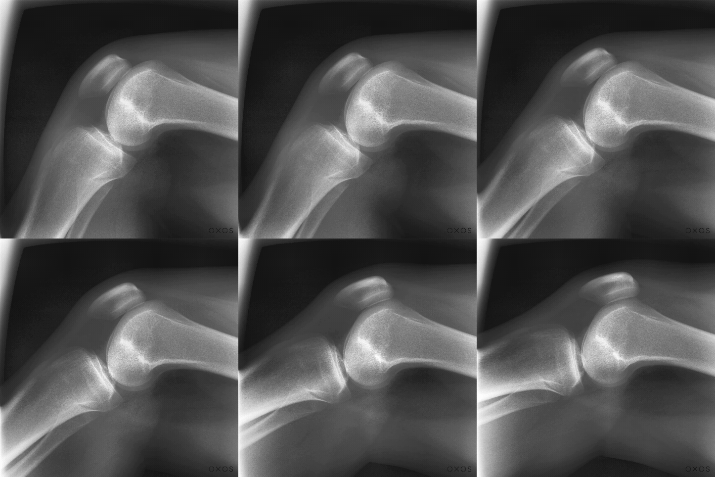
There are a few factors that prevent knee X-rays from being turnkey, despite how often they’re needed. A complete evaluation typically requires multiple projections, each offering a different perspective on the joint. Standard exams include an anteroposterior (AP) view, a lateral view, and a patellofemoral (sunrise) view, increasing the demands on both positioning and workflow.
Those demands are amplified by the wide range of clinical scenarios in which knee imaging is performed. In trauma cases, pain, swelling, or instability may limit a patient’s ability to move. In non-traumatic cases, variability in patient size, mobility, and tolerance for positioning still introduces challenges that can affect consistency and efficiency.
Traditional, room-based X-ray systems often struggle to accommodate this variability. Fixed equipment can make detector placement and patient repositioning time-consuming, particularly when multiple views are required. Technologists are forced to work around physical constraints rather than adapt imaging to the patient in front of them.
When knee X-rays are ordered as frequently as they are, and under such varied conditions, the challenges add up quickly. Small inefficiencies add up. Clinicians see fewer patients over the course of the day, and the patient experience is affected. The combination of imaging requirements, patient limitations, and equipment constraints highlights the need for imaging solutions that can adapt to real-world clinical conditions.
Technique and Versatility Are Critical
Addressing the challenges associated with X-raying the knee starts with technique. Accurate knee imaging depends on obtaining orthogonal views, particularly the anteroposterior (AP) and lateral projections, to fully assess bony anatomy and joint alignment. This requires precise coordination between patient positioning, detector placement, and beam alignment.
In acute injury scenarios, technique becomes even more critical. When patient movement is limited or contraindicated, the horizontal beam lateral (cross-table lateral) view is the preferred approach. With the patient positioned supine, this method minimizes movement of the affected knee while still producing a diagnostically useful lateral image.
Executing these views in practice requires flexibility. The detector must be positioned along the medial aspect of the knee, aligned parallel to the affected leg, while the X-ray beam is directed perpendicular to the long axis of the femur. Stabilization and alignment can be difficult to achieve consistently, particularly outside of traditional radiology suites.
These technical requirements highlight the importance of versatility in knee imaging. X-ray systems should support precise technique while adapting to patient limitations and varied clinical environments. Without that adaptability, even technically straightforward views can become difficult to capture reliably.
MC2 Simplifies Knee X-Rays
MC2 was designed to make X-rays easier, more versatile, and accessible. The portable X-ray device is well-suited for knee X-rays because it adapts to the patient and the clinical scenario, rather than the other way around.
Ease of use
MC2 reduces friction for technologists at every step. Its intuitive setup and maneuverability make it easier to position the system and detector for standard knee projections, including lateral and horizontal beam lateral views. Faster, more straightforward positioning not only improves efficiency but also helps create a smoother experience for patients who may already be in pain or have limited mobility.
Versatility
MC2 is particularly valuable when patient positioning must be adjusted on the fly. Whether imaging is performed with the patient supine, seated, or standing, the system can be positioned to support proper technique without unnecessary repositioning. This flexibility helps maintain consistent image quality across a wide range of clinical scenarios, from acute injuries to planned orthopedic evaluations.
Accessibility
The true portability of MC2 allows knee X-rays to be captured at the point of need, eliminating dependence on dedicated imaging rooms. MC2 fits naturally into orthopedic clinics, inpatient settings, and other care environments where space and time are limited. By enabling efficient capture of standard knee projections without room constraints, MC2 supports both urgent and routine imaging workflows.
Knee X-Ray Imaging Without Compromise
Knee X-rays place unique demands on imaging systems. They require flexibility to accommodate multiple projections, precision to support proper technique, and efficiency to keep workflows moving in high-volume clinical settings.
MC2 supports the full range of knee imaging needs without adding complexity. By combining portability, versatility, and ease of use, MC2 allows orthopedic teams to capture consistent, high-quality knee X-rays across varied clinical scenarios.
The result is a modern approach to one of orthopedics’ most common, and most critical, imaging tasks.
See the MC2 for yourself. Request a demo today.
FAQ
What is a knee X-ray used for?
A knee X-ray is commonly used to evaluate bone structure and joint alignment. Clinicians order knee X-rays to assess suspected fractures after injury, investigate pain or swelling, evaluate joint space narrowing associated with osteoarthritis, and identify signs of infection or joint effusion. Because the knee is a major load-bearing joint, X-rays are often a first-line imaging tool.
What views are included in a standard knee X-ray exam?
A standard knee X-ray exam typically includes multiple views to fully assess the joint. These usually include an anteroposterior (AP) view, a lateral view, and a patellofemoral (sunrise) view. In certain cases, additional views, such as weight-bearing bilateral AP images, may be used to evaluate alignment and joint space under load.
Why is a lateral knee X-ray important in trauma cases?
The lateral knee X-ray provides an orthogonal view to the AP image and is critical for identifying fractures, joint effusions, and alignment issues. In trauma cases, a horizontal beam lateral (cross-table lateral) view is often preferred because it allows imaging with minimal patient movement, reducing discomfort and risk while still providing diagnostic value.
What makes knee X-ray imaging challenging for clinicians and technologists?
Knee X-ray imaging can be challenging. This is due to the need for multiple projections, variability in patient mobility and pain tolerance, and the technical requirements of proper positioning and beam alignment. These challenges are often compounded by fixed, room-dependent equipment, which may limit flexibility in real-world clinical scenarios.